
Station - Instructions for candidate
You are a junior doctor working in the diabetes outpatient clinic of a metropolitan hospital.
Yiannis Georgiou, a 61-year-old musician with type 2 diabetes for 14 years, has been sent to the clinic by the GP for review of poor glycaemic control and six months of gradually blurred vision, worse in the right eye. Fundus photographs were taken by the clinic's retinal camera today and are described for you below, together with the visual acuities and recent results. The patient is sitting with you and would like to know what the pictures show.
Fundus photographs (non-mydriatic retinal camera) and visual acuity
| Item | Value | Reference range |
|---|---|---|
| Visual acuity, right eye | 6/12, no improvement with pinhole | 6/6 |
| Visual acuity, left eye | 6/9, improving to 6/6 with pinhole | 6/6 |
| Left eye: red reflex | Clear | Clear |
| Left eye: disc | Normal colour, margins and cup; no abnormal vessels | Normal |
| Left eye: vessels | Normal calibre; no beading | Normal |
| Left eye: macula | Normal foveal reflex; no deposits within two disc diameters of the fovea | Normal |
| Left eye: periphery | About a dozen tiny red dots and small dot haemorrhages, mostly nasal to the disc; two small clusters of waxy yellow deposits in the far periphery; no fluffy white patches | None |
Recent bloods (GP, 2 weeks ago): HbA1c 9.5% (80 mmol/mol); eGFR 62 mL/min/1.73m2; urine albumin-creatinine ratio 8 mg/mmol; total cholesterol 5.6, LDL 3.4, HDL 1.0, triglycerides 2.8 mmol/L.
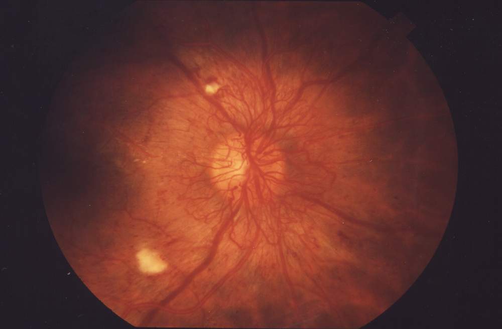
Your tasks are to:
- Interpret the fundus photographs and visual acuities provided.
- Explain what the photographs show to Mr Georgiou.
- Tell Mr Georgiou your diagnosis for each eye and what it means for his sight.
- Discuss the initial management with Mr Georgiou.
- You do NOT need to take a history or examine the patient; the relevant information is provided.
This is a 10-minute station, with 2 minutes of reading time.